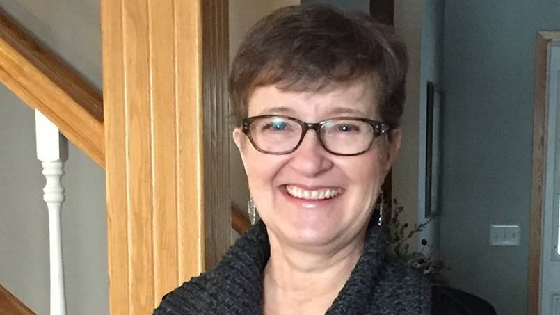
Following cutting-edge brain scanning and precision surgery, Jo Weir is catching up on decades of sleep lost to nighttime seizures, and she's relishing her renewed energy.
After more than 40 years, Jo Marie Weir is sleeping soundly. Plagued by nocturnal seizures that disrupted her nights since she was a teenager, Jo searched for decades to find an accurate diagnosis and effective treatment for her condition.
In 2016, Jo found the answer she was looking for at Mayo Clinic. Neurologists and neurosurgeons used innovative brain imaging, called stereo electroencephalography, to identify and then remove an abnormal portion of her brain that was causing epilepsy.
"I sleep 10 hours a night, and it's so nice," Jo says. "I feel like a different person. I have all this energy, so I went back to work. I also volunteer in an assisted-living facility. At 59 years old, I feel like I finally have a life again."
Coloring Jo's new life is gratitude for the Mayo Clinic specialists who diagnosed her with refractory focal epilepsy in 1995 and, more than 20 years later, succeeded in curing her seizures. The care and treatment Jo received is the epitome of the individualized medicine practiced at Mayo Clinic, says neurosurgeon Jamie Van Gompel, M.D.
"The therapy itself is different and has fewer complications. But it's also the opportunity to have multiple team members give input and evaluate the data that leads to success," Dr. Van Gompel says. "It's very much about the team members planning and working together along with the actual technology."
Exhaustion, ambiguity, despair
Jo was in her first year of college when seizures began disturbing her sleep. "I'd wake up gasping for air," Jo says, adding that during the day, she was sleepy all the time. Jo began seeking answers for her relentless fatigue. In the process, she underwent dozens of electroencephalograms, or EEGs — a test that detects electrical activity in the brain.
"All of the tests that normally show some sort of abnormal brain activity were negative, so nobody believed me," she says.
Despite negative test findings, her doctor prescribed Jo anti-seizure medication. It suppressed her seizures for a few years, during which time she married and had children. But eventually the seizures recurred. In the mid-1990s, Jo went to a family doctor and, through tears, told him she needed to either be seen by a specialist or begin inpatient psychiatric care.
"It's very much about the team members planning and working together along with the actual technology." — Jamie Van Gompel, M.D.
"People don't truly understand what lack of sleep is really like until you've gone through so many years with it," Jo says. "Between lack of sleep and the stress of taking care of three boys and a husband and a household and working part time, it got to be too much."
Jo's pleas compelled her physician to refer her to the Department of Neurology at Mayo Clinic's Rochester campus. Before her appointment, Jo and her husband decided to gather evidence of her seizures.
"My husband didn't want this doctor to think I was crazy, so he videotaped me sleeping," she says.
Diagnosis, validation, medications
At Mayo Clinic, Jo met with neurologist Frank Sharbrough, M.D., now an emeritus professor in the Department of Neurology. Dr. Sharbrough and the Mayo Clinic epilepsy care team presented Jo with the option of undergoing video-electroencephalogram monitoring to record seizures while she was hospitalized. Those recordings showed conclusively that Jo had focal seizures, which most likely originated in the frontal lobe of the brain.
"Of course it was a huge relief," Jo says.
Despite showing the general area of seizure activity, the imaging available at the time couldn't pinpoint the seizure's precise location. That prevented Jo's doctors from planning an effective surgical approach.
Jo had two choices: add additional anti-seizure medications to her regimen or undergo extensive brain surgery. At that time, the technology would have required surgeons to remove a large area of suspected seizure activity. Jo opted to take the medication. For years after that, her seizures were mostly controlled, but in time they came back.
With the return of sleep-disturbing seizures, Jo had a sleep study at a local health care facility. The results were inconclusive about whether seizure activity persisted. She was again referred to Mayo Clinic. Back at Mayo, Jo met neurologist Erik St Louis, M.D. Together they decided she would begin taking different combinations of anti-seizure medication.
"While it is quite scary for some patients to contemplate, epilepsy surgery can lead to seizure control in carefully selected patients who are not seizure-free on at least two different anti-seizure medications." — Erik St Louis, M.D.
"Seizures during sleep are very disruptive to sleep quality and can be dangerous and hard to control in some patients," Dr. St Louis says. "Mrs. Weir was tried intensively on several additional anti-seizure drugs in various combinations for nearly four years."
While two-thirds of epilepsy patients respond to medications, Jo was among the one-third of people who do not.
"We continued to revisit the idea of surgery as possibly being the best option to ultimately control her seizures," Dr. St Louis says.
Eventually Jo began experiencing seizures while awake.
"The development of daytime seizures tipped the balance in her wanting to reconsider brain surgery," Dr. St Louis says. "While it is quite scary for some patients to contemplate, epilepsy surgery can lead to seizure control in carefully selected patients who are not seizure-free on at least two different anti-seizure medications."
MRI, electrodes, breakthrough
The surgical option presented to Jo by her care team — which included Drs. St Louis and Van Gompel, as well as epilepsy specialists Cheolsu Shin, M.D., David Burkholder, M.D., and Lily Wong-Kisiel, M.D. — was a two-step process that began with administering a state-of-the-art brain MRI.
Jo had undergone MRIs previously, but advances in MRI technology through the intervening decades yielded a much different image than Jo's past MRIs. This test revealed a subtle, previously unseen, cortical malformation that Dr. Shin detected after a detailed review of the imaging. The new information allowed Jo's team to consider providing Jo with the more nuanced, targeted approach of stereo electroencephalogram, or sEEG, to further pinpoint her seizure activity.
While invasive brain monitoring is not new at Mayo Clinic, stereo electroencephalograms have only been in practice at Mayo since 2014. Whereas traditional invasive monitoring involves opening the skull (a surgery known as craniotomy) and placing a network of electrodes on the surface of the brain, stereo electroencephalogram allows physicians to bypass a large craniotomy and deliver more electrodes through tiny incisions.
"I was just so excited they could finally see what was going on with me." — Jo Weir
During stereo electroencephalogram, surgeons drill tiny holes in the skull and then insert needle-like electrodes through the holes, deep into the brain tissue. The electrodes, which contain between six and sixteen receptors, allow for the creation of a 3-D representation of the brain.
"The needle-like sEEG approach is more tolerable to patients because we don't have to do a craniotomy," Dr. Wong-Kisiel says. "We can access seizure sites deep in the brain, and we can also introduce these sEEG needles on both sides of the brain, which is much more difficult in an open craniotomy."
In the fall of 2016, eight sets of electrodes were implanted into Jo's brain using stereo electroencephalogram. Then her anti-seizure medicine was stopped, and her team waited for abnormal brain activity that would be tracked via the electrodes.
"The very first night they could see what was going on," Jo says. "I was just so excited they could finally see what was going on with me."
Surgery, recovery, relief
After the electrodes were removed from her brain, Jo went home for six weeks to allow the brain tissue to recover. In November 2016, Dr. Van Gompel, guided by the stereo electroencephalogram information, removed the cortical dysplasia that was responsible for the misfires in Jo's brain.
The first few weeks after surgery were quite difficult. "After I came out of it, I was having a hard time," Jo says. But the treatment she received from her Mayo Clinic team helped mediate her discomfort.
"It was overall, constant care that I was getting," Jo says. "I thought it was fascinating that they had all these people coming from different areas talking to me. During the monitoring, there was a nurse sitting in my room 24 hours a day. I thought it was just amazing that they could see what was going on. It just floored me."
"You hear about this technological stuff, but you just don't expect it to touch your life in some way. It was fascinating, and I am so happy it worked." — Jo Weir
That her seizures have not returned in the year since her surgery and that she's been able to experience fully wakeful days adds to the affinity Jo feels for Mayo Clinic. "Mayo is awesome – the best of the best," she says.
"I have more clarity in my thinking. My memory is better. I do not get tired during the day," Jo says. "I really didn't realize how much the seizure activity affected my ability to function."
Reflecting on her experience, Jo is still intrigued by the technology used to cure her seizures. But most of all, she's just glad the seizures are gone.
"For all those years, they didn't understand it. They finally developed a procedure where they could go in there with the rods and prove the seizure location for removal," she says. "You hear about this technological stuff, but you just don't expect it to touch your life in some way. It was fascinating, and I am so happy it worked."
HELPFUL LINKS
- Read more about epilepsy.
- Learn about the Department of Neurology.
- Connect with others talking about epilepsy and seizures on Mayo Clinic Connect.
- Explore Mayo Clinic.
- Request an appointment.


